
Noninvasive Mapping of the Brain
Physicians use advanced imaging technology to get images of the brain and assist with procedures.
Intraoperative Magnetic Resonance Imaging (iMRI)
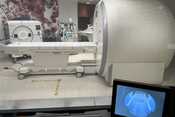 The iMRI is specifically designed to enable magnetic resonance imaging during neurosurgical procedures for brain tumors, epilepsy and other neurological conditions. The iMRI uses a magnetic field to create highly detailed images, enabling doctors to see how the brain is responding and refine the treatment in real-time. Not only is this technology designed to enable greater precision, but also reduces the need for many post-procedure MRIs and no longer requires patients to be moved for imaging during procedures.
MRI-guided focused ultrasound
In addition to the iMRI, the MRI-guided focused ultrasound further expands much needed treatment options in the region. While ultrasound uses sound waves to create images of the body, high-intensity focused ultrasound is a treatment used to ablate or burn targeted tissue. The MRI-guided focused ultrasound uses magnetic resonance images to locate a precise area of tissue and concentrate focused ultrasound energy on the targeted area of the brain, without damaging the surrounding area. This incision-less technique also provides tissue temperature monitoring and real-time patient feedback during treatment. The addition of focused ultrasound to the iMRI makes this a unique environment aimed to improve patient outcomes and offer the most advanced and least invasive way to treat Parkinson’s disease and tremor.
Dr. Zachary Levine, Professor of Neurosurgery and Neurosurgeon at GW Hospital, discusses the MRI-guided focused ultrasound to treat Parkinson's disease and other advanced treatments available at the hospital.
Watch this video to learn more.
Electroencephalogram (EEG)
An EEG provides a record of the electrical activity going on in the brain. A technician connects an EEG machine to specific parts of a patient’s head using wires and electrodes. The electrodes pick up signals from the different parts of the brain and chart those signals as waves that change depending on activity. Epilepsy often produces abnormal brain waves not only during seizures, but also at other times. Physicians in the Epilepsy Center can interpret information from the charts to help make a diagnosis.
Magnetic Resonance Imaging (MRI)
An MRI makes detailed pictures of the brain by using a magnetic field and pulses of radio wave energy. It can detect abnormal blood vessels, tumors, scar tissue, and other lesions that may be responsible for producing seizures.
Positron Emission Tomography (PET)
A PET scan uses a radioactive material called a tracer to highlight areas of concern in the brain.
Single Photon Emission Computed Tomography (SPECT)
Two scans of the brain are performed for this test: one during a seizure and another while the brain is functioning normally. Radioactive material is used to highlight the active sections of the brain and the two scans are compared. The seizure focus (the area that is producing seizures in the brain) is highlighted by subtracting the non-seizure scan from the seizure scan. This test often tremendously helps physicians to make surgical decisions.